Category:Basal-cell carcinoma
Jump to navigation
Jump to search
subtype of basal-cell carcinoma most commonly occurring on the sun-exposed areas of the head and neck  | |||||
| Upload media | |||||
| Instance of |
| ||||
|---|---|---|---|---|---|
| Subclass of |
| ||||
| |||||
Microscopic Pictures - Basal cell carcinoma
Subcategories
This category has only the following subcategory.
H
Media in category "Basal-cell carcinoma"
The following 59 files are in this category, out of 59 total.
-
 517 Basal Cell Carcinoma.jpg 975 × 649; 531 KB
-
 Basal cell carcinoma (1).jpg 1,025 × 683; 92 KB
-
 Basal cell carcinoma (2).jpg 2,700 × 1,800; 2.47 MB
-
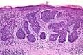
 Basal cell carcinoma Histological diagram.jpg 2,471 × 1,697; 752 KB
-
 Basal Cell Carcinoma left infraorbital cheek.jpg 560 × 240; 32 KB
-
 Basal Cell Carcinoma Left Upper Back nodular and micronodular.jpg 454 × 320; 34 KB
-
 Basal cell carcinoma, nodular.jpg 720 × 480; 53 KB
-
 Basal Cell Carcinoma, Right cheek.jpg 586 × 426; 76 KB
-
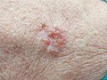
 Basal cell carcinoma, superficial.jpg 720 × 480; 76 KB
-
 Basal Cell Carcinoma, ulcerated, nodular on right lower cheek.jpg 252 × 264; 18 KB
-
 Basal cell carcinoma, ulcerated.jpg 3,000 × 2,000; 627 KB
-
 Basal cell carcinoma.jpg 575 × 383; 54 KB
-
 Basal Cell Carcinoma.png 1,200 × 864; 575 KB
-
 Basal cell carcinoma2.JPG 2,137 × 2,152; 297 KB
-
 Basal cell carcinoma3.JPG 1,473 × 1,220; 278 KB
-
 Basal-cell Carcinoma.jpg 1,023 × 816; 267 KB
-
 Basaliom am Nasenrücken 1.JPG 2,392 × 1,637; 2.6 MB
-
 Basaliom am Nasenrücken 2.JPG 4,752 × 3,168; 6 MB
-
 Basaliom am Nasenrücken 3.JPG 4,752 × 3,168; 5.51 MB
-
 Basaliom am Nasenrücken 4.JPG 4,752 × 3,168; 6.08 MB
-
 Basaliom am Nasenrücken 5.JPG 3,168 × 4,752; 6 MB
-
 Basaliom IMG 5630.JPG 4,272 × 2,848; 3.63 MB
-
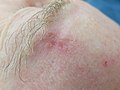
 Basaliom infraorbital rechts.JPG 4,272 × 2,848; 3.57 MB
-
 Basaliom1.jpg 600 × 430; 146 KB
-
 Basaliom2.jpg 600 × 586; 185 KB
-
 Basaliom3.jpg 600 × 487; 166 KB
-
 Basalioma 02.jpg 2,053 × 1,540; 1.94 MB
-
 Basalioma.jpg 2,288 × 1,712; 1.83 MB
-
 BCC Nodular type.jpg 1,200 × 1,818; 610 KB
-
 BCC-mini.jpg 591 × 946; 90 KB
-
 BCC2010.jpg 600 × 909; 155 KB
-
 Cornu cutaneum2.jpg 3,648 × 2,736; 6.66 MB
-
 Dermatoskopie eines nodulären Basalzellkarzinoms, WIKIDERM®.jpg 4,000 × 3,000; 2.88 MB
-
 Dermatoskopie eines pigmentierten Basalzellkarzinom, WIKIDERM®.jpg 4,000 × 3,000; 2.44 MB
-
 Dermatoskopie eines pigmentierten Basalzellkarzinoms.jpg 4,000 × 3,000; 2.87 MB
-
 Dermatoskopie eines superfiziellen Basalzellkarzinoms, WIKIPEDIA®.jpg 4,000 × 3,000; 3.29 MB
-

-
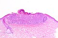
 Histologie eines nodulozystischen Basalzellkarzinoms, WIKIDERM®.jpg 5,184 × 3,456; 1.51 MB
-
 Histologie eines superfiziellen Basalzellkarzinoms, WIKIDERM®.jpg 5,184 × 3,456; 1.7 MB
-
 Histologie eines ulzerierten nodulären Basalzellkarzinoms, WIKIDERM®.jpg 5,184 × 3,456; 1.52 MB
-
 Man with a rodent ulcer on the chin Wellcome L0067009.jpg 3,563 × 5,630; 1.96 MB
-
 Metatypisches Basalzellkarzinom, WIKIDERM®.jpg 4,000 × 3,000; 5.18 MB
-
 Mikronoduläres Basalzellkarzinom, WIKIDERM®.jpg 4,000 × 3,000; 3.9 MB
-
 Noduläres Basalzellkarzinom, WIKIDERM®.jpg 4,000 × 3,000; 4.89 MB
-
 Pigmented BCC.jpg 1,200 × 801; 271 KB
-
 PigmentedBCC.jpg 600 × 400; 79 KB
-
 Präoperative Schnittführung beim nodulären Basalzellkarzinom am Kinn, WIKIDERM®.jpg 3,921 × 2,519; 2.92 MB
-
 Radiography, X-ray therapeutics and radium therapy (1916) (14571777158).jpg 2,269 × 3,679; 681 KB
-
 Scar-like morphea-like BCC.jpg 1,200 × 821; 247 KB
-
 ScarlikeBCC.jpg 600 × 411; 64 KB
-
 Seborrhoeic dermatitis2.jpg 3,648 × 2,736; 6.02 MB
-
 Sklerodermiformes Basalzellkarzinom am Ohr, WIKIDERM®.jpg 4,000 × 3,000; 3.16 MB
-
 Superficial basal cell carcinoma.jpg 4,272 × 2,848; 1.41 MB
-
 Superfizielles Basalzellkarzinom, WIKIDERM®.jpg 4,000 × 3,000; 5.24 MB
-
 The principles and practice of surgery (1916) (14764167865).jpg 1,260 × 1,608; 226 KB
-
 Tumeur basocellulaire.jpg 1,500 × 994; 693 KB
-
 Wisconsin medical recorder (1905) (14595387539).jpg 2,156 × 2,920; 463 KB
-
 Wisconsin medical recorder (1905) (14778902561).jpg 1,976 × 2,540; 767 KB
-
 Wisconsin medical recorder (1905) (14801918213).jpg 2,156 × 2,920; 1.14 MB

.jpg)
.jpg)


_(14571777158).jpg)


_(14764167865).jpg)

_(14595387539).jpg)
_(14778902561).jpg)
_(14801918213).jpg)